Supporting Vulvoscopy
What We Learned at ISSWSH
Seeing More During Vulvoscopy
 At ISSWSH, we are connecting with physicians and advanced practice clinicians who are expanding the conversation around sexual health and taking a closer look at the conditions affecting their patients’ comfort, confidence, and quality of life.
Dr. Sarah Cigna’s presentation on vulvar dermatoses reinforced something that matters deeply to us at Lutech: careful visualization deserves a central place in vulvar care. Her session prompted us to think about how magnified examination, individual patient documentation, and clear communication can support the work these providers are doing.
Every patient has their own anatomy, symptoms, and history. Capturing that individual picture creates a useful reference for the conversations and care that follow.
Looking more closely at persistent symptoms
Itching, burning, irritation, and changes in tissue appearance can have multiple causes. Patients with recurrent symptoms need a careful assessment that considers infection, inflammatory skin conditions, and other explanations.
For suspected fungal infections or vaginitis, images can document visible findings such as redness, swelling, fissures, or discharge when present and accessible during examination. These images record tissue appearance; they do not identify the organism responsible. History, examination, and appropriate testing remain essential, especially when symptoms persist or recur.
For providers, this creates a practical opportunity: document the initial appearance, connect those findings with symptoms and test results, and capture comparable images at follow-up. A visual record can help support a more informed discussion about what has changed.
Helping patients understand what is normal
One of the most meaningful uses of visualization is patient education.
Vestibular papillomatosis, the small fingerlike projections that can appear along the vestibule, is a benign anatomical variation that patients may mistake for genital warts. Visible sebaceous glands, often called Fordyce spots, can also be a normal finding.
When a patient is concerned about a bump or unfamiliar texture, a clinician can offer to show them the area and explain the finding. For a patient who wants to see, that shared view can make the explanation easier to understand and may help reduce anxiety.
The value is in pairing the image with clinical knowledge and a thoughtful conversation. The 2025 AUA/SUFU/AUGS Guideline on Genitourinary Syndrome of Menopause, endorsed by ISSWSH and The Menopause Society, emphasizes individualized clinical evaluation and shared decision-making. As one of the clinical principles states, “Clinicians should educate patients.”
Documenting each patient’s tissue over time
Our notes from Dr. Cigna’s presentation included lichen sclerosus, post-inflammatory pigmentation changes, painful vulvar aphthous ulcers, vulvar Crohn’s disease, and vulvar Paget’s disease. The range of conditions discussed underscored the importance of examining each patient carefully, including the surrounding and perianal tissue when clinically appropriate.
This is where consistent documentation becomes especially valuable. Images can record the location and visible extent of a finding, provide a baseline for future comparison, and add context when another clinician becomes involved.
For patients who need a biopsy or specialist evaluation, including gynecologic oncology when indicated, images can accompany the clinical history and findings. They support communication without replacing an examination, tissue diagnosis, or timely referral.
Making the patient experience part of the care plan
Another takeaway was the importance of comfort during vulvar biopsy.
Discussing pain management, explaining each step, and preparing the patient for recovery deserve deliberate attention. Topical anesthesia, injected local anesthesia, and wound closure when appropriate are among the considerations clinicians can individualize to the procedure and patient.
For us, this reinforced a broader point: patients should understand what is happening and feel that their comfort matters. That experience helps build the trust needed for ongoing care.
How Lutech Supports Vulvoscopy
 As more providers incorporate vulvoscopy into sexual health care, they need practical ways to examine tissue, explain findings, and document what they see.
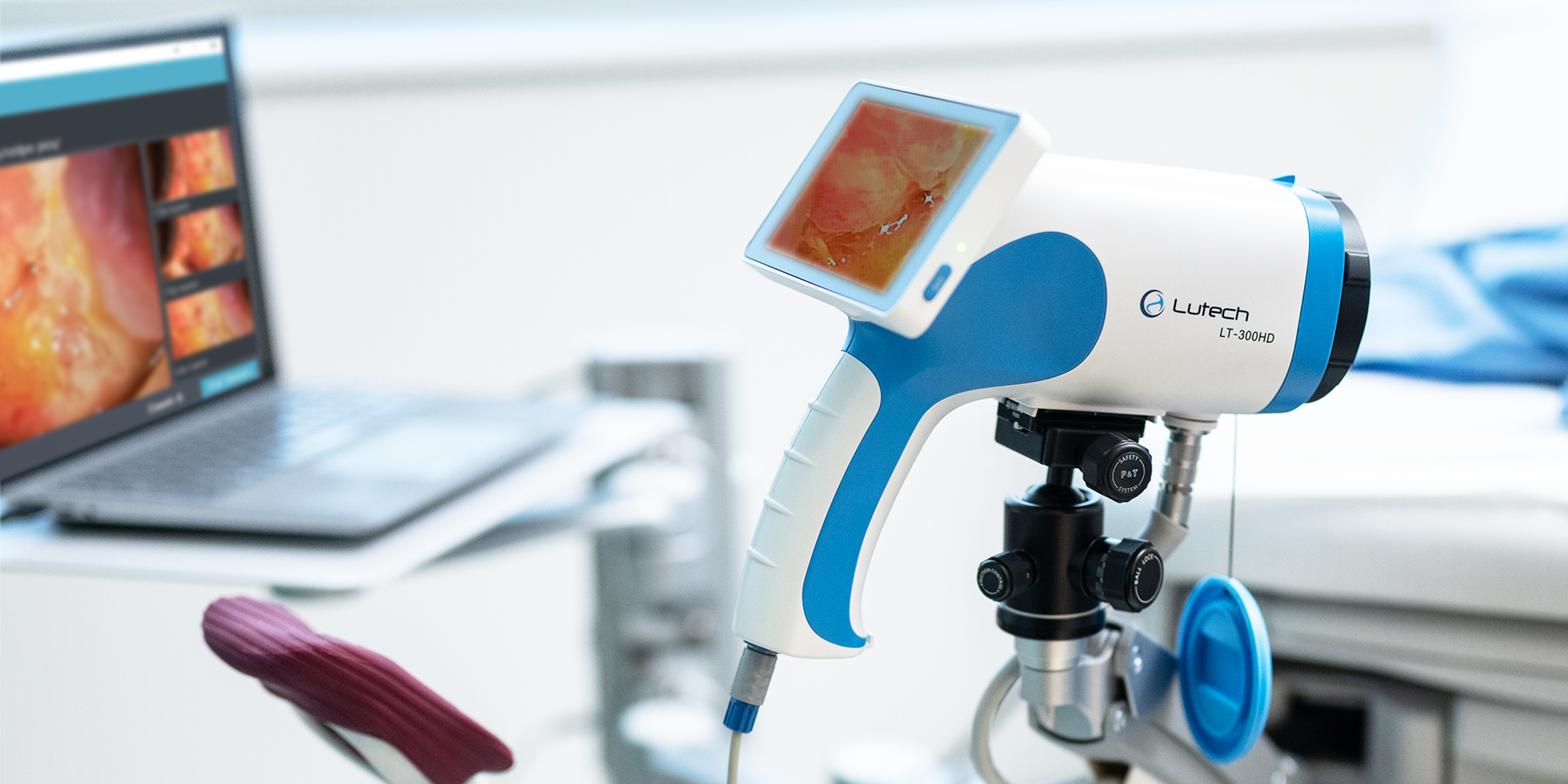
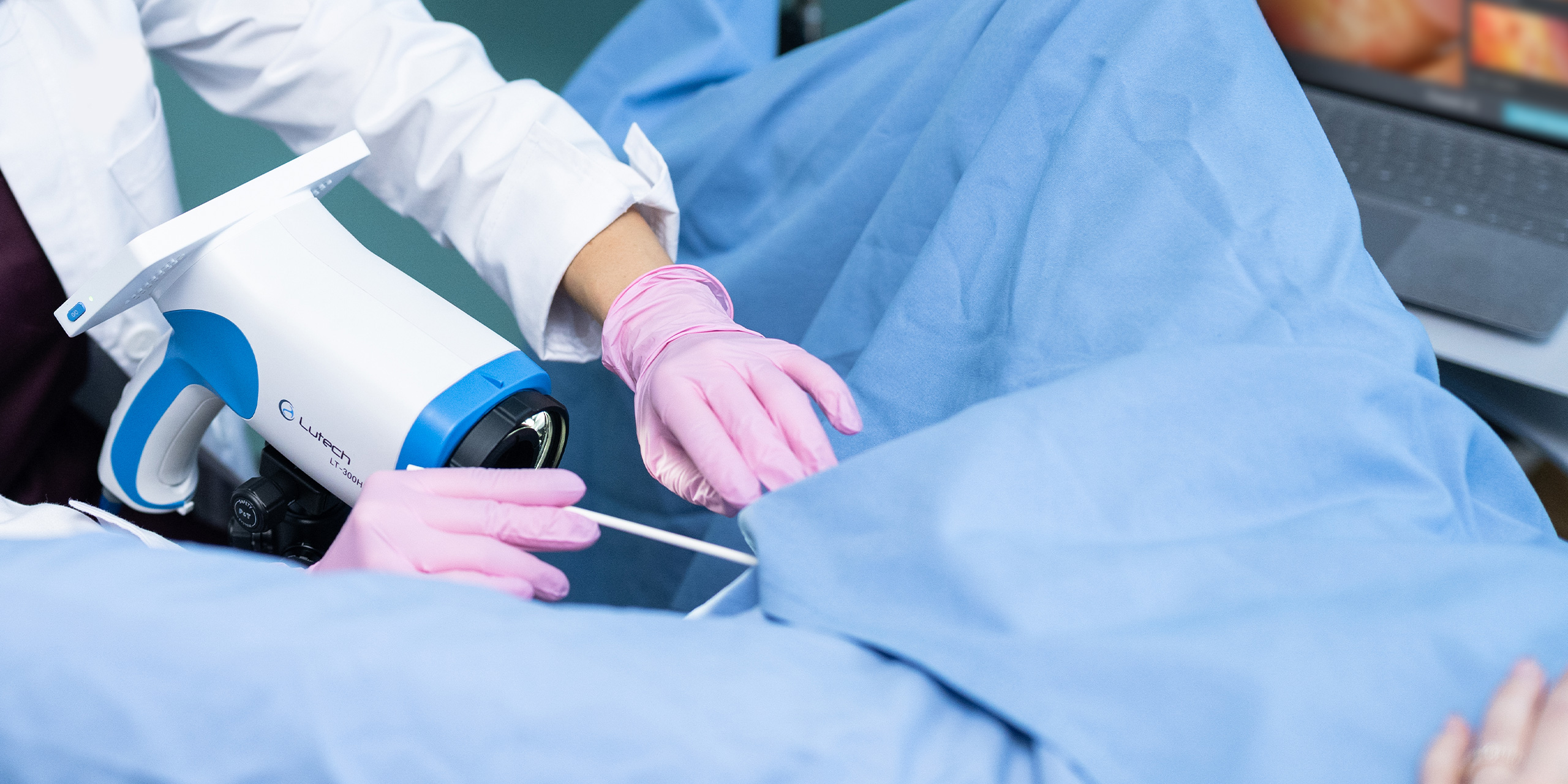
The Lutech LT-300HD brings high-definition digital visualization, optical magnification, and adjustable illumination into that examination workflow. Its digital format supports a shared view of the examination, while image capture can help clinicians build a visual record for follow-up and care coordination.
With patient consent and appropriate secure handling, clinical images can support:
- Baseline documentation of visible tissue findings.
- Comparison across visits.
- Patient education about anatomy and examination findings.
- Communication with referring providers and specialists.
Our role at Lutech is to support the clinicians building more comprehensive sexual health services with technology that fits the way they examine, educate, and care for patients.
ISSWSH is reinforcing how much opportunity there is to improve the vulvar care experience. We are here to listen, learn, and help providers put clearer visualization and meaningful documentation into everyday practice.
Interested in incorporating digital vulvoscopy into your practice?
Connect with Lutech and request a demonstration of the LT-300HD today.
AUA/SUFU/AUGS. (2025). Guideline on Genitourinary Syndrome of Menopause. Endorsed by ISSWSH and The Menopause Society.